Balancing the Normal Foot: Hoof Preparation, Shoe Fit and Shoe Modification in the Performance Horse (Part 1)
Part 1 references are at the end of part 2, which will appear in the October issue of Anvil Magazine.)
© O.K. Balch, D. Butler and M.A. Collier
published in ANVIL Magazine, September 1998
Reprinted with permission from Equine Veterinary Education, 1997
Equine Sports Medicine Laboratory and Comparative Orthopaedic Research Laboratory, Department of Veterinary Medicine and Surgery, College of Veterinary Medicine, Oklahoma State University, Stillwater, Oklahoma 74078-0107 and Butler Publishing and Farrier Services, PO Box 1390, Laporte, Colorado 80535, USA
INTRODUCTION
Appropriate hoof preparation and shoe fit are integral to soundness and performance. (6, 7, 10) Historically, inappropriate modification (trimming) and extension (shoeing) of the hoof capsule have been considered the root cause of foot pathology. (17, 41) Currently, diseases of the foot are considered the predominant cause of equine lameness. (25)
The overall relationship between farriery and the ability of the limb (and thus the horse) to absorb concussion and to function efficiently and safely is compelling and obvious. A cohort study of 95 Thoroughbred horses concluded that appropriate trimming and shoeing could reduce musculoskeletal disease and enhance performance. (21) A case-control study of Thoroughbred horses that were euthanized on California racetracks identified toe grabs on aluminum racing plates as significant risk factors in fatal musculoskeletal injuries associated with suspensory apparatus failures and metacarpal/metatarsal (cannon bone) condylar fractures. (20)
Appropriate hoof balance requires an appreciation of the conformation and athletic activity of the horse. Within the context of this paper, appropriate hoof balance is defined as hoof preparation that enhances performance and interferes minimally with long-term athletic ability. Stricter attention to specific details of trimming and shoeing yields greater dividends for the equine athlete due to increased musculoskeletal demands inherent to performance. Important elements of foot preparation include the following:
- hoof angle
- hoof length
- mediolateral hoof orientation
- thickness of the sole, frog, and bar
- wall contour and ground surface (3)
Shoes function as an important component of hoof balance by adding length and weight to the digit. Shoes also alter the ground-contact area of the hoof, its frictional characteristics, and its relative position to the axis of the limb.
This review of hoof preparation, shoe fit, and shoe modification provides specific guidelines that may be useful for performance horses. Conformation and intended use are the final determinants for the appropriateness of specific trimming and shoeing techniques for an individual horse.
HOOF PREPARATION
Hoof angle
Hoof angle is the dorsal to solar angulation of the hoof measured at the toe. Hooves are described as broken forward or club footed (high hoof angle) when the angle of the hoof is too steep for its associated pastern; conversely, hooves are described as broken backward (low hoof angle) when the angle of hoof is too sloping for its associated pastern. (Fig. 1) (6) Classical and modern renditions of hoof flight patterns portray a uniphasic (single-humped) parabolic curve with the shape and often the length of the curve determined by hoof angle (Fig. 2) (17, 39, 41, 45).
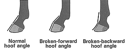 Fig. 1: Absence of dorsal alignment of the hoof and pastern creates broken-forward and broken-backward hooves. |
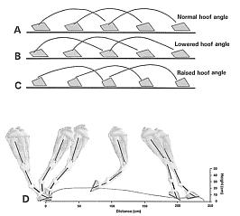 Fig. 2: Hoof-flight patterns: Presumed and actual. Drawings A, B, and C are adapted from A. Dollar's 1898 classic depiction of the effects of hoof angle alterations on hoof flight. These drawings were not based on cinematographic studies. Unfortunately, these quite inaccurate basic patterns are still displayed in current textbooks. Drawing A represents the presumed hoof flight that is produced by a normal hoof angle and characterized by a uniphasic parabolic curve with its maximal height centered on the parabolic curve. Drawings B and C display the presumed effects that either lowering or raising the hoof angle would shift the point of maximal height of the parabolic curve either caudally or cranially, respectively. Cinematographic and videographic studies of equine locomotion have demonstrated definitively that hoof flight patterns are biphasic; the first maximum which occurred shortly after toe off is always higher than the second maximum, which occurred shortly before initial ground contact. Drawing D represents seven limb positions in a stride representing 6 horses trotting at 4 m/s and shod with normal angle and hoof length. (Balch et al 1996) |
Altering the angle of the hoof affects limb kinematics and kinetics through predictable alterations in the suspensory apparatus (Fig. 3). Hyperextension (excessive overextension) of the metacarpo (metatarso) phalangeal (fetlock) joint is limited by the suspensory apparatus of the fetlock, a specialized portion of the stay apparatus. The suspensory apparatus in the forelimb consists of the following anatomic structures: the deep digital flexor tendon and its associated accessory (inferior check) ligament; the superficial digital flexor tendon and its associated accessory (superior check) ligament; the suspensory ligament with its dorsal branches and its continuation the distal sesamoidean ligaments; and the paired proximal sesamoids. The suspensory apparatus of the hindlimb is functionally similar, though slight differences exist anatomically. Collectively, the suspensory apparatus of both the forelimb and hindlimb forms a ligamentous "hammock" which cradles the fetlock during the stance phase. Energy is stored in the elastic suspensory apparatus while the fetlock moves to its most distal and overextended position at the mid-stance position. (16) After the limb moves beyond the mid-stance position, the release of stored energy assists the flexion of the distal limb at lift off and the elevation of the fetlock. (11) Logically, a change in the tension of one of the three structures that comprise the suspensory apparatus should result in a compensatory change in the tension of the other structures or a change in the relative degree of overextension of the fetlock or both.
 Fig. 3: Effects of hoof angle modifications on the joints, tendons, and ligaments of the distal portion of the forelimb. Raising the hoof angle flexes the distal interphalangeal joint, flexes the proximal interphalangeal joint slightly, and extends the metacarpophalangeal joint very slightly. Lowering the hoof angle has the opposite effects on these joints. Raising the hoof angle decreases tension in the deep digital flexor tendon and increases tension in the superficial digital flexor tendon. Lowering the hoof angle increases tension in the deep digital flexor tendon. (Adapted with permission from Balch et al., 1995) |
Altering the hoof angle changes the relative position of the flexor surface of the distal phalanx and, subsequently, the tension of the deep digital flexor tendon which inserts at that location. Not surprisingly, lowering the hoof angle (either by trimming heel or adding a toe wedge) increased maximal tension in that tendon in walking horses. (28, 40) Raising the hoof angle (either by trimming the toe or adding a heel wedge) decreased maximal tension. (28, 40, 46) Conversely, when the hoof angle was raised, the mean maximal tension in the superficial digital flexor tendon and the suspensory ligament increased. (46) However, the changes in the superficial digital flexor tendon were only significant at the trot. (40, 46) Radiographic studies have demonstrated that lowering the hoof angle extends the distal interphalangeal (coffin) joint, extends the proximal interphalangeal (pastern) joint slightly, and flexes the fetlock joint very slightly. Conversely, raising the hoof angle produced the opposite effects on the joints of the distal limbs. (9)
Despite the popularity of the often-portrayed rendition of hoof flight as an uniphasic parabolic curve influenced by hoof angle changes, a cinematographic study (12) demonstrated that hoof flight was more accurately depicted as a biphasic (double-humped) parabolic curve (Fig. 2) with the hoof angle exerting little effect on shape or length of the hoof-flight pattern. Additionally, low or high angles did not affect stride length in horses walking, trotting, and cantering on a high-speed treadmill. (4) A low hoof angle slowed (increased) breakover, the period between heel off and toe off; (4, 12) a high hoof angle quickened (decreased) breakover. (4)
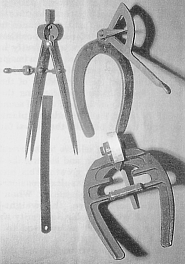
Fig. 4: Hoof protractors, calipers, and rulers are used to measure quantitatively hoof angle and hoof length. |
A hoof is trimmed appropriately when the dorsal surfaces of the hoof and pastern are parallel. (3, 14, 38) Visual confirmation requires that horses stand squarely on all limbs with cannon bones positioned vertically. Use of a hoof protractor quantifies the hoof angle and facilitates record keeping of the hoof angle of individual hooves over time. (Fig. 4) Successful use of a protractor requires that the distal two-thirds of the hoof wall be dressed with a rasp so that it is a straight continuation of the proximal third (without dishes or flares).
Studies using instrumented shoes on horses moving on a treadmill at a walk, trot, and canter determined that forces at the toe, medial heel, and lateral heel were collectively the lowest when the hoof and pastern regions were aligned. (5) This parallelism occurs for most horses when fore hooves are trimmed between 50 and 54 degrees and hind hooves between 53 and 57 degrees. (3) Conformation of the lower portion of the limb, especially the pastern, may vary substantially with breed and individual. (9) Pastern variation may also occur between paired limbs of a horse; horses afflicted with unilateral chronic suspensory desmitis often experience contracture of the suspensory apparatus which extends the fetlock joint and increases the slope of the pastern in the affected limb. (6) The angle of the hoof should be related to the conformation of that limb. (3)
HOOF LENGTH
Hoof length is the measurement of the length of the hoof wall. The hoof is most commonly measured on the dorsum of the wall from the center of the toe to the coronary rim, the most proximal portion of the wall. Careful palpation of the coronet, the junction between the skin and the wall, reveals the most proximal extent of hoof wall, which feels hard in contrast to the soft cutaneous tissue covering the distal aspect of the pastern. The border between the hoof and the hair on the pastern is approximately the same level as the coronary rim and is readily recognized unless the hair at the coronet is allowed to grow long. The use of calipers or rulers, either indirectly or directly, provides quantitative measurements. (Fig. 4) Accurate measurements of hoof length are important to owners and trainers since many breed and show associations enforce restrictions on maximum hoof length. (27) For example, the regulations of the American Horse Shows Association specify a maximum toe length for horses competing in Arabian and Morgan divisions. (27) The Horse Protection Regulations issued by the United States Department of Agriculture to eliminate soaring abuses in Tennessee Walking Horses and racking horses limit the height of pads to no more than half the length of the natural hoof wall (Horse Protection Act of 1970).
| Table 1 - Guidelines for hoof length based on the weight of the horse | ||||
| Horse Weight | Toe Length | |||
| Horse Size | Kg | Pounds | Cm | Inches |
| Small | 360 - 400 | 800 - 900 | 7.6 | 3.0 |
| Medium | 425 - 475 | 950 - 1050 | 8.25 | 3.25 |
| Large | 525 - 575 | 1150 - 1250 | 8.9 | 3.5 |
| These guidelines apply to most breeds of nongaited horses; however exceptions occur. If the horse will be barefoot in a turnout environment, an additional 0.5 to 0.6 centimeter (3/16 to 1/4 inch) of wall and a thicker sole is advisable. | ||||
Lengthening the hoof increases the length of the lever arm over which the hoof pivots at the end of the stance phase. A videographic study investigated the effects of augmented hoof length (2.5 and 5.0 cm) in trotting horses. (5) The effects were four-fold on the kinematics of the hoof. Lengthening the hoof:
- increased the maximum height of the hoof during the biphasic hoof flight
- increased the vertical velocity of the hoof
- shortened the horizontal distance between toe off and the achievement of maximal height of the hoof
- slowed breakover.
Despite traditional expectations of farriers and trainers, lengthening the hoof did not affect stride length. (5) Lengthening the hoof amplifies torque on the distal portion of the limb and increases pressure on the navicular bursa by the deep digital flexor tendon at the end of the stance phase. This increased length of the lever arm may lead to injuries of the suspensory apparatus. (36) As the hoof grows and the toe moves away from the axis of the coffin joint, the tendency of the horse to stumble, display awkward gaits, and become injured by limb contact increases. (3)
Hoof length, either excessively short or long, may cause lameness. (6) When trimming and shoeing short-footed (nongaited) horses, guidelines (Table 1) for hoof length can be based on the weight of the animal, with the exception of racing Standardbreds. (3) Racing Standardbreds are shod to maximize trotting or pacing speed. Trotters generally require more carpal flexion (folding action) than pacers and are often shod with longer hooves. (43) Horses of other breeds that compete with an animated trot are shod similarly.
MEDIOLATERAL HOOF ORIENTATION
Mediolateral hoof orientation is determined by the relative lengths and angles of the medial and lateral walls of the hoof capsule and is then modified to achieve mediolateral balance for the limb. Two different techniques are used to judge how to trim one side of the hoof relative to the other. (3)
The first technique, and the most commonly used, is a geometric limb-axis-oriented procedure that prepares the ground surface of the wall perpendicular to the axes of the cannon bone and phalanges. (Fig. 5) Variations include the use of other anatomic structures as guidelines such as the hairline at the coronet, the bulbs of the heels, the axis of the phalanges, or change in medial or lateral orientation of the digit as it is manually flexed, extended, and overextended at the fetlock.
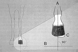 Fig. 5: Attaining mediolateral balance through the commonly used geometric-limb-axis-oriented technique. Line A represents the axes of the cannon bone and phalanges. The wedge labeled C is the portion of the hoof necessary to be trimmed if the solar surface of the hoof is to be made perpendicular to line A. (Adapted with permission from Balch et al 1991) |
The second technique is result-directed and requires that hoof-landing patterns be viewed. Commonly, the hooves are trimmed so that hooves land simultaneously on the medial and lateral heels or land simultaneously on all segments of the bearing surface of the hoof (flat-footed). The more the horse's limb conformation approaches that of the ideal horse (conformation characterized by limb straightness, proper angulation of joints, and a stance that is neither base-wide or base-narrow), the more likely the two different techniques will result in similarly trimmed hooves. Conversely, the more the horse's limb exhibits conformational deviations, the less likely the two techniques will result in similarly trimmed hooves. For example, if the mediolateral aspect of the hooves of horses possessing a base-narrow, toe-out forelimb conformation is trimmed perpendicular to the axes of the cannon bone and phalanges, the hooves will commonly land lateral-heel first as opposed to flat-footed. (Fig. 6)
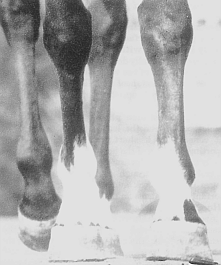 Fig. 6: Attaining mediolateral balance through result-directed trimming. The left front hoof lands first on the lateral aspect of the wall. Result-directed trimming dictates that the lateral side of the hoof be trimmed shorter so that the medial and lateral heels land simultaneously. (Adapted with permission from Balch et al 1991) |
Instrumented-shoe studies examining horses exercising on a high-speed treadmill and measuring force on the toe, medial heel, and lateral heel characterized weight distribution across the solar surface of the hoof during the stance phase and hoof-landing patterns at various gaits. (5) Contrary to common expectations, hooves that landed flat-footed or simultaneously on the medial and lateral heels did not evenly distribute force across the solar surface of the wall, but rather the medial side of the hoof was preferentially weighted during the stance phase. The hoof-landing patterns were primarily lateral-heel first in freshly trimmed horses that were judged visually by two experienced farriers as landing simultaneously on both heels or flat-footed at a walk. Instrumented shoes, measuring ground contact in milliseconds, identified variations in hoof-landing patterns that were not distinguishable to unaided human vision, because the events that defined the landing patterns occurred at intervals shorter than the temporal resolution of human vision. Also, the same study indicated that the landing patterns observed at a walk did not necessarily reflect those landings at a trot or canter.
Achievement of appropriate mediolateral orientation is challenging in well-conformed horses and may be very difficult in the performance horse with conformational deviations. Which of the two general techniques, geometric-limb-axis-oriented procedure or result-directed procedure, that should be used in an individual horse is often controversial. In the sound performance horse, a geometric limb-axis-oriented procedure using the least distorted anatomy as a guideline is the most efficient technique. When the horse is lame and the source of lameness is located within the digit, trimming the solar surface of the hoof to achieve simultaneous landing of both heels may effectively treat the lameness in many cases and in most cases is a useful therapeutic supplement to more specific veterinary treatment. Traditionally, lamenesses associated with inappropriate mediolateral balance and identified as lateral distorted hooves, (17, 30) sheared heels, chronic heel soreness, navicular syndrome, (32) and chronic metacarpophalangeal synovitis (2) have been treated by trimming to achieve simultaneous landings of the medial and lateral heel.
THICKNESS OF THE SOLE, FROG AND BAR
The solar surface of the hoof capsule consists of the distal projection of the wall and horny layers of the sole, frog, and bars. These horny (epidermal) layers cover and protect their associated coria (dermis) from trauma. Thicknesses of these epidermal structures are difficult to measure directly in the intact horse but can be estimated radiographically. Approximately 6 to 9 mm (1/4 to 3/8 inch) of horny sole covers sole corium in average-sized Quarter Horses; (10) the sole is thinner in Thoroughbreds and Warmbloods.
Weight bearing on a limb affects sole descent and heel expansion during the stance phase. As early as 1810, Clark correlated the flattening of the sole concavity and abaxial expansion of the heels and concluded that interference with normal heel expansion would be detrimental to soundness. (37) Experimental studies done by Lungwitz (1891) and Dollar (1898) concluded that thickness of the sole, as well as width of the hoof, affects sole descent and heel expansion when the limb is weight bearing. The presence of the circumflex artery and vein of the sole and a large palmar/planter (solar) venous plexus sandwiched between the horny sole and the distal surface of the third phalanx predisposes the foot to bruising if the sole is used for more than minimal weight bearing. The insensitive sole acts primarily as a protective covering for deeper sensitive tissue.
The frog has a limited weight-bearing function, although regular ground contact is thought to stimulate normal frog growth and heel expansion. (3) Traditional theories relate frog pressure to heel expansion. Accepted by many veterinarians and farriers, the frog pressure/heel expansion tenet has been theorized as ground pressure on the frog being distributed proximally into the digital cushion and then passed abaxially through the hoof cartilages and venous plexes to cause outward movement of the wall at the heels and quarters. (10)
Results of recent experimental studies suggest that the relationship between frog pressure and heel expansion is more complex. Colles (1989) directly evaluated the relationship between frog pressure and hoof wall expansion using foil strain gauges attached to the wall. In this study, all horses and ponies with normal frog pressure displayed heel expansion, however, reduced frog pressure resulted in heel contraction in some horses and ponies, but heel expansion in others. Enhanced frog pressure resulted in even less consistent results with heel expansion in some horses, contraction in others, and in some, expansion and contraction depending on the stage of weight bearing, as well as lameness in some animals. In the opinion of the authors, periodic weighting of the frog during normal locomotion stimulates frog growth and promotes heel expansion in most horses; however, the geometry of the hoof capsule plays a significant role in determining individual response.
Unshod horses regularly wear away the outer insensitive sole and bars as those structures lose moisture, and becomes flaky. This normal exfoliation is accompanied by distinct molting of the frog semi-annually. (3) Shoes diminish the normal abrasiveness of the ground and subsequent thinning of these epidermal structures. Superfluous sole, frog, and bars should be trimmed. The sole should be thinned so that it is concave and not in the same plane as the ground surface of the wall; ideally, the ground surface of the sole should be trimmed to parallel the curved solar surface of the distal phalanx so that uniform thickness is maintained. However, excessive thinning of the sole predisposes the underlying sole dermis to bruising. The sole at the toe can usually be thinned until glossy or "live" sole is visible. (10) Manual palpation of the sole is useful in estimating its thickness; the degree of springiness is related to sole thickness or distance to the underlying corium. (3)
If the heels of the hoof are normally shaped and not contracted, the frog may be trimmed level with the ground surface of the finished wall. Anaerobic organisms (such as Fusobacterium necrophorum) are commonly found in the collateral and central sulci of the frog, but rarely cause disease unless trapped in air-deprived recesses. Removal of ragged edges and loose pieces of frog decreases these pockets that may harbor the organisms causing thrush. (10) The collateral sulci should be regularly cleaned and opened, especially adjacent to the widest part of the frog, to allow escape of debris that collects routinely in these natural recesses. The bars can be trimmed below the level of the ground surface of the finished wall and contoured to match the concaved adjacent sole. Excessive trimming of the bars is discouraged, as bars may act as internal struts to inhibit heel contraction in the absence of normal frog pressure. (3)
WALL CONTOUR AND GROUND SURFACE
Wall contour is the circumferential shape of the wall, and the ground surface is the portion of the wall directly in contact with the ground. Wall contour and ground surface partially determine the size of the hoof and structural stability of the wall. Ground-contact surface of the hoof (or its extension of the shoe) positions the weight-bearing surface of the limb in specific spatial relationships to the rest of the limb. During the stance phase, the fetlock acts as an articulation between the loaded fore (hind) limb and the digit. The digit consists of a short link (the pastern formed by the first and second phalanges) articulating with a supporting platform (the hoof enclosing the distal phalanx) which is always positioned dorsally to force vectors associated with the horse's weight and transmitted through cannon bones. Both angular changes in the fetlock and deformation of the hoof are important mechanisms for dissipation of concussive forces.
Hoof size reflects the physical dimensions of the enclosed distal phalanx and is determined by the angle, contour, thickness, and length of the wall at the toe, quarters, and heels, the prominence and shape of the frog and bars, and the thickness of the epidermal sole and frog. Accurate assessment of hoof size requires that the walls of hoof be dressed (rasped) so that the distal two-thirds is a straight continuation of the proximal third of the wall. A 1988 study of farriery management of navicular syndrome identified the hoof characteristic of a small-foot-to-body-weight as statistically more common in horses afflicted with navicular syndrome than a matched group of sound horses. (48) Certain trimming and shoeing techniques have been incriminated in the development of unnecessarily small feet. Shoes that are inadequately sized for the hoof, nailing behind (palmar/plantar) to the widest part of the hoof, and excessive trimming of the frog and bars hinder the physiological expansion of the horse's heels during the stance phase. These techniques promote heel contracture. In some horses and breeds, the hoof characteristic of a small-foot-to-body size may be an inheritable trait and difficult to modify. (48)
Slightly different configurations of the body, limbs, and hooves have been described as ideal for dissipation of concussion and maintenance of soundness. (1, 17, 19, 44) In 1903, Russell presented empirically derived guidelines for identifying ideal limb-to-hoof conformation and specified with geometric precision that the "normal center of equipoise" is located slightly caudal to the apex of the frog. (41) Force-plate studies, measuring the horizontal and vertical forces exerted by hooves and conducted 75 years later, confirmed that the point of zero moment (location of a single vector representing all the complex forces applied to the digit) was centered on the cranial third of the frog at the walk and trot. (42)
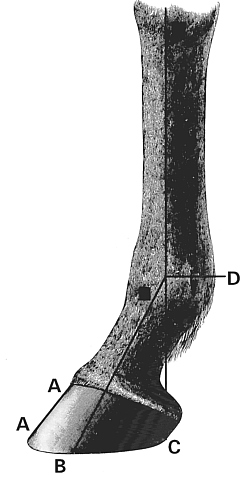 Fig. 7: Relative position of the hoof in relationship to the rest of the limb. Line B represents the "angle of incidence" defined by the axes of the phalangeal bones. Line C bisects the metacarpus (metatarsus) and extends distally to brush the palmar (plantar) border of the heels on the ground surface of the wall. (illustration adapted from Russell, W. 1903) |
Russell's drawings (Fig. 7) provide useful guidelines for the relative position of the hoof (shoe) in relationship to the rest of the limb. The "normal center of equipoise" is the termination of an "angle of incidence" passing distally through the axes of the phalanges and identifies the center of the hoof. Russell specified that the distance from this center point to any circumferential position on the hoof (shoe) must be equal to secure a perfectly balanced foot. Most importantly, he specified that an imaginary line passing distally from the center of the fetlock joint should brush the most palmar (plantar) extent of the ground surface of the wall. Further characterizing the normal palmar (plantar) surface of the hoof wall of sound horses, Murray in 1873 stated that the palmar (plantar) face of the hoof wall should parallel the dorsal face of the hoof wall.
The position of the hoof relative to the overall axis of the limb influences trimming and shoeing the hoof and subsequent direction of hoof growth. It becomes an especially important biomechanical consideration in horses with angular limb deformities. When viewed from a cranial perspective, the weight-bearing surface of a limb with angular deformity should be centered under the fetlock as opposed to centered under the hoof capsule. (Fig. 8)
 Fig. 8: Use of a shoe to extend the ground-bearing surface of a limb with angular deformities. Illustration A depicts a weanling with multiple limb angular deformities: mild carpal valgus with lateral rotation and severe fetlock varus with medial rotation. The angular deformity of the fetlock displaces the force vectors that are associated with the horse's weight laterally, relative to the ground surface of the hoof; the upright, heavily loaded lateral heel and the flare of the wall at the medial toe are consequences. Illustration B uses red vertical lines adjacent to the medial and lateral borders of the fetlock to suggest the appropriate ground support for the limb. Illustrations C and D depict the appropriately trimmed hoof and the trimmed and shod hoof with a lateral extension, respectively. As suggested by the contours of the fetlock, osteoarthritis was confirmed radiographically. Shoeing with a lateral extension was palliative, not curative. |
The weight of the horse is transmitted through the skeleton to the distal phalanx, which in turn is suspended from the inner surface of the hoof wall by the stratum internum (the interdigitation of the epidermal and dermal laminae). The hoof wall and, to a lesser extent, the bars, are well designed for weight bearing as the stratum medium, the bulk of the hoof wall and bars, is composed of hoof tubules. Hoof wall derives strength and shape from the orderly proximodistal arrangement of tubular and intertubular horn. Strength is compromised when hoof tubules in the toe and quarter regions bend abaxially as they near the ground surface and create a dished or flared hoof wall. In the normal horse, hoof tubules in the medial and lateral heels are straight and parallel those in the dorsal hoof wall. If the palmar/plantar surface of the hoof slopes more than that of the dorsal wall, then the load on the most distal portion of the palmar/plantar hoof wall is increased. This increased force may collapse the hoof wall. The palmar/plantar hoof wall continues to lose its parallelism to the dorsal hoof wall as the tubular and intertubular horn realigns itself to the ground surface.
This loss of parallelism causes diminished palmar (less commonly plantar) ground support, is very common in performance horses, and is referred to as underrun heels, sloping heels, (10) collapsed heels, (14) underslung heel, (34) or run-under heels when the palmar face diverges at least 5 degrees from dorsal face. (48) The presence of underrun heels may not be synonymous with disease in the limb, but is a strong biomechanical predisposition to injury. A 1988 study of 50 sound performance horses identified that 26 of the horses had underrun heels. (48) The prevalence of underrun heels in racing Thoroughbreds and Quarter Horses (35) supports the contention that this hoof conformation may be an inherited characteristic that predisposes horses to chronic heel soreness and other musculoskeletal injuries. The hoof tubules in the portion of the heel that is underrun are bent forward, diminishing their ability to resist compression. Concussion is disproportionately concentrated in the heels due to the simultaneous reduction in ground surface area and dorsal shift in location. This dorsal shift of ground-contact surface hyperextends the coffin joint, increases tensile forces on the palmar (plantar) aspect of the limb, and increases compressive loading of the dorsal surface of the limb. (51)
When viewed from a cranial (caudal) perspective, the ground surface of the hoof should be centered under the axis formed by radius and metacarpus (tibia and metatarsus) in well-conformed horses. When viewed laterally, an imaginary line that bisects the cannon bone should intersect the most palmar (plantar) extent of the ground surface of the wall. The distal two thirds of the wall at the toe, quarters, and heels should be a straight continuation of the proximal third of the wall. The palmar (plantar) surface of the wall should parallel the dorsal surface. Shoes function as continuations of hoof capsules. Shoeing can extend the ground surface of the limb to compensate for ill-shaped hooves (e.g., underrun heels) and poorly conformed limbs.
Part 1 references are at the end of part 2, which will appear in the October issue of Anvil Magazine.)
Return to the Farrier Articles listing page.
Return to the ANVIL Online Table of Contents for September, 1998.